Kirsch operator
Edge detector
The Kirsch operator or Kirsch compass kernel is a non-linear edge detector that finds the maximum edge strength in a few predetermined directions. It is named after the computer scientist Russell Kirsch.
Mathematical description
The operator takes a single kernel mask and rotates it in 45 degree increments through all 8 compass directions: N, NW, W, SW, S, SE, E, and NE. The edge magnitude of the Kirsch operator is calculated as the maximum magnitude across all directions:

where z enumerates the compass direction kernels g:
- and so on.


The edge direction is defined by the mask that produces the maximum edge magnitude.
Example images
-
 Original
 -
 Maximum gradient in the 8 directions


-
 Image filtered with g(1)
 -
 Image filtered with g(2)
 -
 Image filtered with g(3)
 -
 Image filtered with g(4)


-
 Image filtered with g(5)
 -
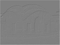 Image filtered with g(6)
 -
 Image filtered with g(7)
 -
 Image filtered with g(8)


References
- Kirsch, R. (1971). "Computer determination of the constituent structure of biological images". Computers and Biomedical Research. 4 (3): 315–328. CiteSeerX 10.1.1.161.956. doi:10.1016/0010-4809(71)90034-6.


