Suboccipital nerve
| Suboccipital nerve | |
|---|---|
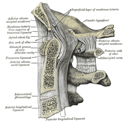 Median sagittal section through the occipital bone and first three cervical vertebrae. (Suboccipital nerve labeled at center right.) | |
| Details | |
| Innervates | Rectus capitis posterior major, rectus capitis posterior minor, obliquus capitis superior, obliquus capitis inferior |
| Identifiers | |
| Latin | nervus suboccipitalis |
| TA98 | A14.2.02.007 |
| TA2 | 6365 |
| FMA | 7054 |
| Anatomical terms of neuroanatomy [edit on Wikidata] | |
The suboccipital nerve (first cervical dorsal ramus) is the dorsal primary ramus of the first cervical nerve (C1). It exits the spinal cord between the skull and the first cervical vertebra, the atlas.[1]
It lies within the suboccipital triangle along with the vertebral artery, where the artery enters the foramen magnum.
It supplies muscles of the suboccipital triangle, the rectus capitis posterior major, obliquus capitis superior, and obliquus capitis inferior. The suboccipital nerve also innervates rectus capitis posterior minor.
See also
Additional images
-
 Upper part of medulla spinalis and hind- and mid-brains; posterior aspect, exposed in situ.
 -
 Suboccipital triangle


References
- ^ Standring, Susan (2016). Gray's anatomy: the anatomical basis of clinical practice (41 ed.). Elsevier Limited. pp. 762–773. ISBN 978-0-7020-5230-9.
| Authority control databases |
|
|---|
 | This neuroanatomy article is a stub. You can help Wikipedia by expanding it. |
- v
- t
- e


