| AKT1 |
|---|
|
| Доступные структуры |
|---|
| PDB | Поиск ортологов: PDBe RCSB |
|---|
| Список идентификаторов PDB |
|---|
1H10, 1UNP, 1UNQ, 1UNR, 2UVM, 2UZR, 2UZS, 3CQU, 3CQW, 3MV5, 3MVH, 3O96, 3OCB, 3OW4, 3QKK, 3QKL, 3QKM, 4EJN, 4EKK, 4EKL, 4GV1, 5KCV |
|
|
| Идентификаторы |
|---|
| Псевдонимы | AKT1, AKT, CWS6, PKB, PKB-ALPHA, PRKBA, RAC, RAC-ALPHA, AKT serine/threonine kinase 1 |
|---|
| Внешние ID | OMIM: 164730 MGI: 87986 HomoloGene: 3785 GeneCards: AKT1 |
|---|
|
|
| Паттерн экспрессии РНК |
|---|
| Bgee | | Человек | Мышь (ортолог) |
|---|
| Наибольшая экспрессия в | - stromal cell of endometrium
- left adrenal cortex
- right adrenal cortex
- ventricular zone
- body of stomach
- muscle layer of sigmoid colon
|
| | Наибольшая экспрессия в | - ventricular zone
- efferent ductule
- endothelial cell of lymphatic vessel
- stroma of bone marrow
|
| | Дополнительные справочные данные |
|
|---|
| BioGPS | 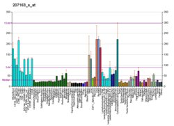 | | Дополнительные справочные данные |
|
|---|
|
| Генная онтология |
|---|
| Молекулярная функция | - GTPase activating protein binding
- kinase activity
- nitric-oxide synthase regulator activity
- АТФ-связанные
- protein kinase activity
- protein phosphatase 2A binding
- enzyme binding
- phosphatidylinositol-3,4,5-trisphosphate binding
- трансферазная активность
- 14-3-3 protein binding
- связывание с белками плазмы
- protein serine/threonine/tyrosine kinase activity
- protein kinase binding
- protein kinase C binding
- нуклеотид-связывающий
- phosphatidylinositol-3,4-bisphosphate binding
- связывание похожих белков
- protein serine/threonine kinase activity
- гомодимеризация белка
- calmodulin binding
| | Компонент клетки | - цитоплазма
- цитозоль
- мембрана
- cell-cell junction
- митохондрия
- клеточное ядро
- ciliary basal body
- microtubule cytoskeleton
- клеточная мембрана
- веретено деления
- нуклеоплазма
- везикула
- постсинапс
- protein-containing complex
| | Биологический процесс | | | Источники: Amigo, QuickGO |
|
| Ортологи |
|---|
| Вид | Человек | Мышь |
|---|
| Entrez | | |
|---|
| Ensembl | | |
|---|
| UniProt | | |
|---|
| RefSeq (мРНК) | NM_001014431
NM_001014432
NM_005163
NM_001382430
NM_001382431
|
|---|
NM_001382432
NM_001382433 |
| |
|---|
NM_001165894
NM_009652
NM_001331107 |
|
|---|
| RefSeq (белок) | NP_001014431
NP_001014432
NP_005154
NP_001369359
NP_001369360
|
|---|
NP_001369361
NP_001369362 |
| |
|---|
NP_001159366
NP_001318036
NP_033782 |
|
|---|
| Локус (UCSC) | Chr 14: 104.77 – 104.8 Mb | Chr 12: 112.62 – 112.64 Mb |
|---|
| Поиск по PubMed | Искать[3] | Искать[4] |
|---|
|
 Информация в Викиданных Информация в Викиданных |
| Смотреть (человек) | Смотреть (мышь) |
|
AKT1 (англ. RAC-alpha serine/threonine-protein kinase, Protein kinase B alpha) — внутриклеточный фермент, один из трёх членов семейства протеинкиназ B. Киназа AKT1 является ключевым ферментом сигнального пути PI3K/AKT и вовлечена в регуляцию пролиферации, роста и выживания клеток[5][6]. Исследованию функций этого фермента уделяется большое внимание из-за того, что он выступает в роли онкогена при многих злокачественных заболеваниях[7]. Ген AKT был впервые клонирован из генома онкогенного ретровируса в 1987 году. Тогда же было обнаружено, что существует и клеточная форма этого гена[8].
Структура
Киназа AKT1 состоит из 480 аминокислотных остатков. По своей структуре она очень близка к двум другим членам семейства протеинкиназ B, AKT2 и AKT3, и включает следующие домены: N-концевой домен гомологичный плекстрину (англ. pleckstrin homology (PH) domain), киназный домен и C-концевой регуляторный домен, содержащий гидрофобный мотив. Регуляция активности киназы происходит за счёт фосфорилирования/дефосфорилирования двух аминокислотных остатков — Thr-308 в киназном домене и Ser-473 в гидрофобном мотиве[9].
Роль в передаче сигнала
AKT1 является важным компонентом фосфатидилинозитол-3-киназного (PI3K) сигнального пути, она имеет множество субстратов и способствует передаче сигнала по этому пути. Большую часть времени AKT1 находится в клетке в неактивном состоянии. При активации PI3K-сигнального пути в клетке синтезируется вторичный мессенджер фосфатидилинозитол-3,4,5-трифосфат (PI(3,4,5)P3). Плекстрин-подобный домен AKT1 связывается с PI(3,4,5)P3, в результате чего киназа перемещается в плазматическую мембрану. Здесь она фосфорилируется ферментами PDK1 (англ. 3-phosphoinositide-dependent protein kinase 1) по остатку Thr-308 и mTORC2 (англ. mammalian target of rapamycin complex 2) или DNA-PK (англ. DNA-dependent protein kinase) по положению Ser-473. Такое фосфорилирование активирует AKT1, она покидает мембрану и перемещается в цитоплазму и ядро клетки, где фосфорилирует белки-мишени, которые обеспечивают дальнейший клеточный ответ. Инактивация киназы осуществляется за счёт её дефосфорилирования под действием фосфатаз PP2 (англ. protein phosphatase 2) and PHLPP (англ. PH domain leucine-rich repeat phosphatase)[9].
Субстраты AKT1
На сегодняшний день известно более 100 субстратов AKT. Фосфорилирование киназой AKT может иметь разные последствия для белка-мишени, включая активацию, инактивацию, изменение внутриклеточной локализации и стабильности. Долгое время считалось, что все субстраты являются общими для трёх изоформ киназы, однако новые данные говорят о том, что, скорее всего, имеет место определённая специализация изоформ по функциям. Так, считается, что AKT1, главным образом, фосфорилирует белки, отвечающие за выживание и рост клеток (MDM2, BAD, p21CIP1, p27KIP1, mTOR и др.)[9].
В некоторых исследованиях ген AKT1 ассоциирован с шизофренией[10].
Примечания
- ↑ 1 2 3 GRCh38: Ensembl release 89: ENSG00000142208 - Ensembl, May 2017
- ↑ 1 2 3 GRCm38: Ensembl release 89: ENSMUSG00000001729 - Ensembl, May 2017
- ↑ Ссылка на публикацию человека на PubMed: (неопр.) Национальный центр биотехнологической информации, Национальная медицинская библиотека США.
- ↑ Ссылка на публикацию мыши на PubMed: (неопр.) Национальный центр биотехнологической информации, Национальная медицинская библиотека США.
- ↑ Chen W. S., Xu P. Z., Gottlob K., Chen M. L., Sokol K., Shiyanova T., Roninson I., Weng W., Suzuki R., Tobe K., Kadowaki T., Hay N. Growth retardation and increased apoptosis in mice with homozygous disruption of the Akt1 gene // Genes Dev.. — 2001. — Т. 15, вып. 17. — С. 2203—2208. — PMID 11544177. Архивировано 26 мая 2016 года.
- ↑ Cho H., Thorvaldsen J. L., Chu Q., Feng F., Birnbaum M. J. Akt1/PKBalpha is required for normal growth but dispensable for maintenance of glucose homeostasis in mice // J Biol Chem.. — 2001. — Т. 276, вып. 42. — С. 38349—38352. — PMID 11533044. Архивировано 9 июля 2017 года.
- ↑ Chin Y. R., Toker A. Akt isoform-specific signaling in breast cancer: uncovering an anti-migratory role for palladin // Cell Adh Migr.. — 2011. — Т. 5, вып. 3. — С. 211—214. — PMID 21519185.
- ↑ Staal S. P. Molecular cloning of the akt oncogene and its human homologues AKT1 and AKT2: amplification of AKT1 in a primary human gastric adenocarcinoma // Proc Natl Acad Sci U S A.. — 1987. — Т. 84, вып. 14. — С. 5034—5037. — PMID 3037531.
- ↑ 1 2 3 Hers I., Vincent E. E., Tavaré J. M. Akt signalling in health and disease // Cell Signal.. — 2011. — Т. 23, вып. 10. — С. 1515-1527. — PMID 21620960.
- ↑ Gene Overview of All Published Schizophrenia-Association Studies for AKT1 Архивировано 21 февраля 2009 года. — база данных SZgene сайта Schizophrenia Research Forum